

Supracondylar Fracture Of The Elbow: Avoiding Misreadings In Pediatrics
Orthopedic surgeons specializing in pediatrics work to minimize the risk of misreadings on x-rays. Supracondylar fracture of the elbow is a common diagnosis in children and misreadings can result in unnecessary treatment and delay recovery. Tips for avoiding misreadings for Supracondylar fracture of the elbow are detailed in this article from Dr. TD Long, creator of MedRev, the only healthcare reputation management system built for healthcare professionals, by healthcare professionals.
3 view elbow x-ray without abnormal rotation is necessary for optimal evaluation.
The lateral view should be performed in 90 degree flexion without rotation. The frontal and oblique views should be performed in fully extended position without overlap of the joint.
Incorrectly positioned elbow views are one of the most common reasons fractures are not seen. The humeral heads should be superimposed on the lateral view with neither head appearing more anteriorly or posteriorly positioned to the other.
When standard views are not performed, the incorrectly positioned view or study should be repeated. With acute injuries of the elbow, optimal views are sometimes just not possible. If the views are not diagnostic, follow-up the study, or a CT of the elbow may be necessary.
Are Your X-ray Technologist Performing The Correct Views Without Rotation Or Override?
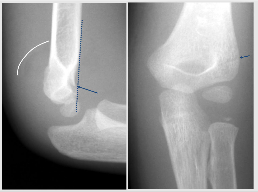
This type of fracture is usually seen with immature patients and unfused epiphyses. The above images demonstrate the pertinent findings.
The left side image demonstrates a large joint effusion which is best noted posteriorly. The white curved line shows the border of the effusion. The anterior humeral line passes abnormally through the anterior one-third of capitellar epiphysis. The normal anterior humeral line usually passes through the mid part of the capitellar epiphysis.
The right side image demonstrates a subtle transverse fracture line through the distal humeral head. The fracture is also seen on the lateral view marked by the blue arrow on the left side image.
Supracondylar fractures are very common in children from a fall on an outstretched arm. These fractures may be very subtle with the fracture line not seen initially. The earliest finding may be joint effusion only.
Joint effusions
Joint effusions can have multiple causes but are most common with trauma. Effusions are usually due to fracture, ligamentous injury, cartilaginous injury or a combination. Persistent severe pain is usually associated with significant injury to the joint or bone. Follow up x-rays in a few days to a week after injury may visualize a fracture. CT scanning can immediately visualize subtle fractures. MRI is necessary for ligamentous or cartilaginous injury.
Proper treatment of supracondylar fractures is necessary for anatomical alignment. However, a large number of these fractures are not displaced and need only immobilization by cast or splint.
Are you looking to improve your patient experience, get better online reviews, and grow your practice?
Learn how MedRev can get you the data and reputation improvement you need to grow.